
Researchers at UCL and the Francis Crick Institute have, for the primary time, recognized the origin of cardiac cells utilizing 3D photographs of a coronary heart forming in real-time, inside a dwelling mouse embryo.
For the examine, revealed in The EMBO Journal, the workforce used a method referred to as superior light-sheet microscopy on a specifically engineered mouse mannequin. It is a technique the place a skinny sheet of sunshine is used to light up and take detailed photos of tiny samples, creating clear 3D photographs with out inflicting any injury to dwelling tissue.
By doing this, they have been capable of monitor particular person cells as they moved and divided over the course of two days—from a important stage of improvement generally known as gastrulation by means of to the purpose the place the primitive coronary heart begins to take form. This allowed the researchers to establish the mobile origins of the guts.
Gastrulation is the method by which cells start to specialize and arrange into the physique’s major constructions, together with the guts. In people, this happens through the second week of being pregnant.
The examine’s findings might revolutionize how scientists perceive and deal with congenital coronary heart defects, the researchers say.
Senior creator Dr. Kenzo Ivanovitch (UCL Nice Ormond Avenue Institute of Youngster Well being and British Coronary heart Basis Intermediate Analysis Fellow) mentioned, “That is the primary time we have been capable of watch coronary heart cells this intently, for this lengthy, throughout mammalian improvement. We first needed to reliably develop the embryos in a dish over lengthy intervals, from just a few hours to some days, and what we discovered was completely sudden.”
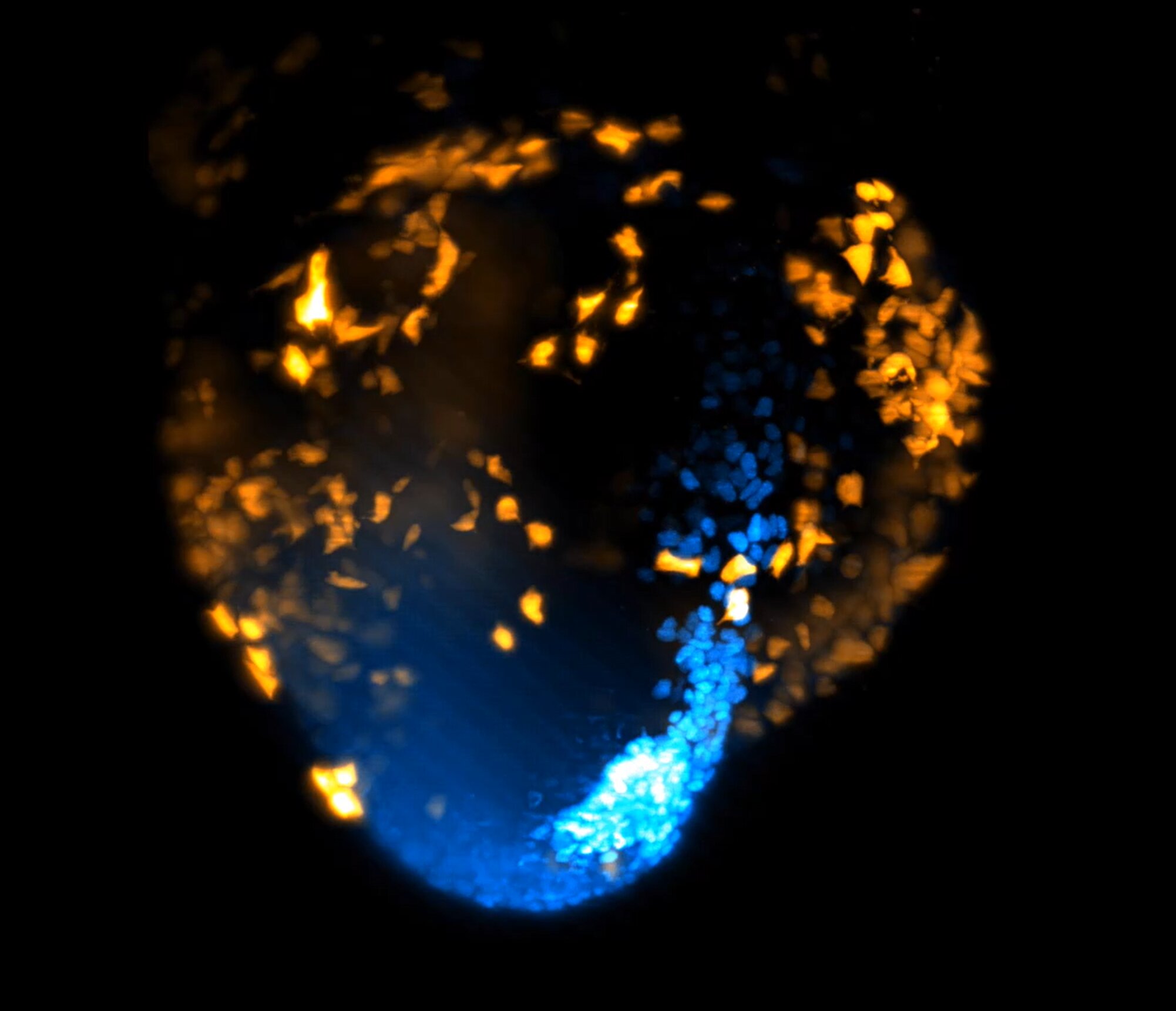
Utilizing fluorescent markers, the workforce tagged coronary heart muscle cells (referred to as cardiomyocytes), inflicting them to glow in distinct colours. Mixed with light-sheet microscopy, this innovation allowed the researchers to create an in depth time-lapse video.
Snapshots have been captured each two minutes over 40 hours, producing photographs with unprecedented spatial decision.
The ensuing footage confirmed how cells transfer, divide, and type the primary elements of an embryo, like the guts. Every glowing cardiomyocyte might then be tracked again to earlier cells, permitting scientists to create a household tree of the cells. This helped them see precisely when and the place the primary cells that solely make the guts appeared within the embryo.
On the very earliest phases, embryonic cells have been multipotent (able to turning into varied cell varieties). These included not solely coronary heart cells but in addition others comparable to endocardial cells, a kind of cell that strains the inside surfaces of blood vessels and coronary heart chambers.
Nevertheless, the researchers discovered that early throughout gastrulation (usually throughout the first 4 to 5 hours, after the primary cell division), cells contributing solely to the guts emerge quickly and behave in extremely organized methods.
Fairly than transferring randomly, they comply with distinct paths—nearly as in the event that they already know the place they’re going and what position they may play, whether or not contributing to the ventricles (the guts’s pumping chambers) or the atria (the place blood enters the guts from the physique and lungs).
Dr. Ivanovitch mentioned, “Our findings display that cardiac destiny willpower and directional cell motion could also be regulated a lot earlier within the embryo than present fashions counsel.
“This basically modifications our understanding of cardiac improvement by displaying that what seems to be chaotic cell migration is definitely ruled by hidden patterns that guarantee correct coronary heart formation.”
Lead creator, Ph.D. candidate Shayma Abukar (UCL Nice Ormond Avenue Institute of Youngster Well being and UCL Institute for Cardiovascular Science) mentioned, “We at the moment are working to know the alerts that coordinate this advanced choreography of cell actions throughout early coronary heart improvement.
“The guts does not come from a single group of cells, it types from a coalition of distinct cell teams that seem at totally different occasions and locations throughout gastrulation.”
The insights from the examine might revolutionize how scientists perceive and deal with congenital coronary heart defects, which have an effect on almost one in 100 infants. The findings might additionally speed up progress in rising coronary heart tissue within the lab to be used in regenerative drugs.
Dr. Ivanovitch mentioned, “Sooner or later, we hope this work will assist uncover new mechanisms of organ formation. This can inform design ideas to exactly program tissue patterns and shapes for tissue engineering.”
Extra data:
Shayma Abukar et al, Early coordination of cell migration and cardiac destiny willpower throughout mammalian gastrulation, The EMBO Journal (2025). DOI: 10.1038/s44318-025-00441-0
Quotation:
Scientists movie the guts forming in 3D sooner than ever earlier than (2025, Could 13)
retrieved 13 Could 2025
from https://medicalxpress.com/information/2025-05-scientists-heart-3d-earlier.html
This doc is topic to copyright. Other than any truthful dealing for the aim of personal examine or analysis, no
half could also be reproduced with out the written permission. The content material is offered for data functions solely.