A 69-year-old lady was referred to the retina service for irregular posterior phase findings.
The affected person’s medical historical past was notable for bilateral listening to loss and kind 2 diabetes recognized 10 years in the past. Whereas her HbA1c peaked to 12.7% 2 years prior, it has since remained steady at 5.8% on dulaglutide, empagliflozin and metformin. She moved from Vietnam to the US 40 years in the past. In any other case, she had no different notable social or household historical past.

Supply: Ke Zeng, MD, Jonathan Caranfa, MD, PharmD, and Marisa Tieger, MD
The view to her posterior phase was beforehand restricted by cataracts. After cataract surgical procedure, her ophthalmologist despatched a referral to retina given the severity and asymmetry in retinal hemorrhages.
Examination
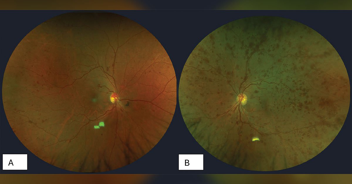
Greatest corrected visible acuity was 20/25 in the fitting eye and 20/20 within the left eye. IOP, pupillary examination, furtherocular actions and confrontational visible fields had been all regular bilaterally. Anterior phase examination was unremarkable in each eyes apart from delicate indicators of dry eye illness and posterior chamber IOLs bilaterally. There was no neovascularization of the iris. A dilated fundus examination revealed shunt vessels on the optic disc in the fitting eye. Each eyes had microaneurysms. The left eye had diffuse dot-blot hemorrhages in all 4 quadrants (Determine 1). Each eyes had dilated and tortuous veins. An inferior horseshoe tear with pigment and no subretinal fluid was famous within the left eye.
Further imaging
Macula OCT confirmed retinal thickening and hint quantities of cystic fluid within the left eye (Determine 2). Fluorescein angiogram confirmed intensive peripheral nonperfusion and diffuse pinpoint microaneurysms in each eyes with a traditional transit (Determine 3).


What’s your prognosis?
See reply beneath.
Irregular posterior phase
Extreme nonproliferative diabetic retinopathy was on the prime of this affected person’s differential prognosis. Retinal vein occlusion was thought of because of the severity of retinal hemorrhages and presence of shunt vessels on the optic disc, nevertheless it was dominated out with regular transit on fluorescein angiogram. Attributable to discordance within the severity of retinal hemorrhage with the affected person’s total well-controlled diabetes, extra workup was pursued with hematology.
Workup and administration
Hypercoagulability workup, which included PT/INR, apolipoprotein B, cardiolipin antibodies, issue VIII exercise, issue V Leiden, useful protein C/protein S exercise, protein C resistance and serum protein electrophoresis, was regular. Antithrombin III ranges had been mildly elevated.


A whole blood depend confirmed a imply corpuscular quantity (MCV) of 64.7 fL, in step with microcytosis. Pink blood depend (RBC) was elevated at 5.89 million/µL with elevated reticulocytes. Mentzer index (MCV divided by RBC) of 11 prompt the affected person’s anemia was extra doubtless associated to thalassemia than iron deficiency, which might extra doubtless current with Mentzer index above 13. A peripheral blood smear and hemoglobin electrophoresis later confirmed beta thalassemia minor.
Dialogue
Thalassemia is an inherited blood dysfunction affecting manufacturing of the alpha and/or beta subunits of hemoglobin. Beta thalassemia minor happens when a mutation happens in one among two beta-globin genes. Systemically, it will probably current with delicate anemia and related signs of shortness of breath, fatigue and dizziness. In beta thalassemia main, mutations happen in each beta-globin genes. That is related to extra extreme anemia and signs akin to jaundice, bone abnormalities and spleen enlargement.
Quite a few ocular findings associated to beta thalassemia have been cited within the literature. This consists of cataracts, anterior ischemic optic neuropathy, retinal pigment epithelium degeneration and angioid streaks. Different posterior findings embody retinal venous tortuosity, retinal hemorrhage, and retinal vein and arterial occlusion. Even proliferative retinopathy has been documented in sufferers with beta thalassemia minor. Beta thalassemia with its inherent hypoxic and hypercoagulable state can place sufferers in danger for ischemic ocular situations.
One may hypothesize that mixed thalassemia and diabetes, each situations with options of ischemia, would exacerbate retinopathy. Counterintuitively, Incorvaia and colleagues confirmed a potential protecting impact of thalassemia by discovering much less extreme retinopathy in sufferers with each thalassemia and diabetes in contrast with these with solely diabetes. There stays vital potential for additional analysis on this space.
In sufferers with each thalassemia and diabetes, it’s value contemplating different strategies for glucose monitoring apart from HbA1c. HbA1c values can turn out to be artificially suppressed in thalassemia because of excessive crimson blood cell turnover. An alternate marker to contemplate is fructosamine, a measurement of principally glycated albumin, which might not be affected in sufferers with thalassemia.
Sufferers with thalassemia are additionally in danger for iron overload because of ineffective erythropoiesis and administration, typically involving blood transfusions. In iron-overloaded sufferers, microaneurysms showing as diabetic retinopathy have been reported. Notably, deferoxamine, an iron chelator used together with blood transfusions, may trigger ocular toxicity. This consists of retinal pigment epithelium modifications related to lowered imaginative and prescient, night time blindness or visible discipline impairments.
Scientific course
The affected person developed macular edema within the left eye with imaginative and prescient worsening to twenty/40. After two intravitreal injections of bevacizumab, her imaginative and prescient improved to twenty/25 with practically full decision of intraretinal fluid. The retinal hemorrhages improved in the fitting eye and remained steady within the left eye. Her hematologist really helpful that she begin each day folic acid supplementation and keep away from iron-containing dietary supplements to stop iron overload.
- References:
- Azimi A, et al. BMC Ophthalmol. 2025;doi:10.1186/s12886-025-04028-5.
- Bhoiwala DL, et al. Surv Ophthalmol. 2016;doi:10.1016/j.survophthal.2015.08.005.
- Hudson JR. Br J Ophthalmol. 1953;doi:10.1136/bjo.37.4.242.
- Incorvaia C, et al. J Pediatr Endocrinol Metab. 1998;11 Suppl 3:879-883.
- Li H, et al. Retin Circumstances Temporary Rep. 2024;doi:10.1097/ICB.0000000000001392.
- Stultz RD, et al. Ophthalmic Surg Lasers Imaging Retina. 2018;doi:10.3928/23258160-20181002-22.
- For extra info:
- Edited by William W. Binotti, MD, and Julia Ernst, MD, PhD, of New England Eye Heart, Tufts College College of Drugs. They are often reached at william.binotti@tuftsmedicine.org and julia.ernst@tuftsmedicine.org.