Up to date on January 29, 2025
Glaucoma is a gaggle of eye ailments with totally different signs, however the frequent function amongst all forms of glaucoma is optic nerve harm. Very like Alzheimer’s illness is a neurodegenerative illness of the mind, glaucoma is taken into account a neurodegenerative dysfunction of the optic nerve.
In glaucoma, optic nerve cells degenerate and ultimately result in cell loss of life, which might trigger everlasting imaginative and prescient loss. This will happen due to elevated eye strain, poor blood movement, and lots of different attainable elements.
Be taught extra about how scientists all over the world are learning methods to regenerate the optic nerve.
What’s the Optic Nerve?
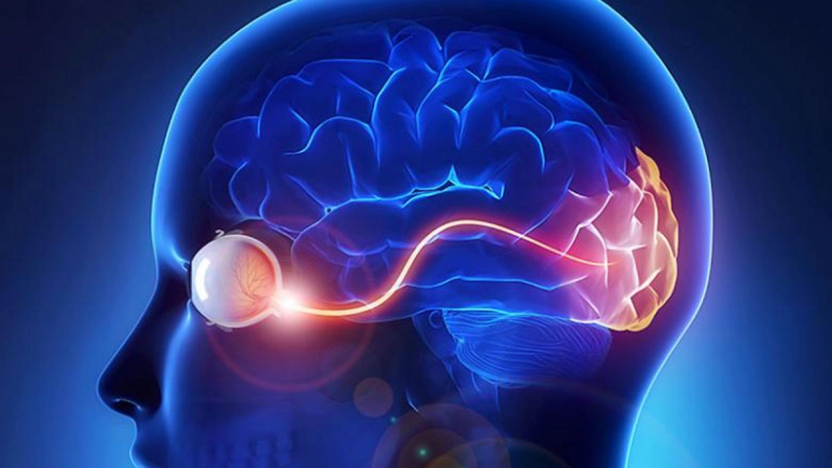
The optic nerve consists of roughly 1.5 million nerve fibers behind the attention that carry visible messages from the retina to the mind. When mild hits the retina, the photoreceptors (light-sensitive cells) obtain and transmit this data to different specialised cells, together with the ultimate cell kind within the chain, known as the retinal ganglion cells. These cells reside within the retina, however their output “cables” or “fibers,” known as axons, prolong from the optic nerve to particular areas within the mind. Retinal ganglion cells play a vital function because the output nerve cell of the attention that transmits visible data to the mind. Within the mind, the visible data is additional processed to provide us sight.
Throughout a watch examination, your eye physician can visualize the optic nerve with the assistance of particular lenses. The axons of the optic nerve are bundled and situated at the back of the attention. This “optic disc” is seen at the back of the attention together with blood vessels. In optic nerve degeneration associated to glaucoma, the optic disc shows modifications which can be attribute of glaucoma, which your physician could check with as “cupping.”
Video Displaying the Optic Nerve
“Cupping”
The traditional optic nerve has a wholesome showing “rim” of tissue, which is assessed by each the contour of the rim in addition to the colour. “Cupping” is the results of modifications within the optic nerve associated to optic nerve degeneration, the place there’s a backward bowing of the central a part of the disc. When your optic disc is seen in three dimensions, the “cupping” could be very apparent to your eye physician. Within the video beneath, the part starting at roughly 29 seconds demonstrates the cupping impact.
Different bodily options of the optic disc can counsel glaucoma, corresponding to thinning of the “rim,” focal notches or lack of rim tissue, and bleeding (additionally known as disc hemorrhages). The thinning of the rim that characteristically first impacts areas of the optic disc in glaucoma is said to the standard visible area deficits seen in glaucoma sufferers.
When “cupping” associated to glaucoma could be very extreme (when it impacts a lot of the rim of the disc such that little or no wholesome tissue stays, for instance), lack of central imaginative and prescient and ensuing blindness may end up, though this usually happens within the very late levels of glaucoma.
Video Demonstrating the “Cupping” Impact
A wonderful visualization of optic nerve “cupping” begins at roughly 29 seconds into the video.
What Occurs to the Optic Nerve in Glaucoma?
In glaucoma, the axons of those optic nerve cells degenerate, and ultimately result in cell loss of life. There are a lot of various factors that contribute to the method. One main danger issue is eye strain. Nonetheless, it’s attainable to have elevated eye strain, or ocular hypertension, and present no modifications of optic nerve degeneration (though one would have to be monitored over time for indicators of degeneration). Conversely, it’s attainable to have regular eye strain and important optic nerve degeneration, which is commonly known as “regular pressure glaucoma” or “low pressure glaucoma.”
What we do know is that there’s possible mechanical harm on the website the place the optic nerve inserts into the again of the attention. It is because the axons of the optic nerve go away the attention by passing by a construction known as the lamina cribrosa, a mesh-like construction composed of pores or holes by which the axon bundles should cross. The location of preliminary harm often is the axons as they cross by the lamina cribrosa because the optic nerve exits the attention.
One other contributing issue could also be blood movement to the optic nerve. Some researchers consider that in instances of normal-tension glaucoma, impaired blood movement to the optic nerve could play a extra essential function within the degenerative course of.
Sadly, like different central nervous system areas such because the spinal twine, the regenerative capability of the optic nerve is proscribed. There are a lot of causes for this, together with the truth that retinal ganglion cells can not restore or regenerate themselves after harm with out important assist.
The surroundings during which the optic nerve resides additionally comprises indicators that inhibit regeneration and lacks indicators to stimulate regrowth. Due to this fact, glaucoma is at present an incurable illness. Nonetheless, scientists all over the world are learning methods to regenerate the optic nerve, not solely to assist glaucoma sufferers, but additionally as a result of the optic nerve is especially useful in learning central nervous system regeneration.
Can the Optic Nerve Regenerate?
Regeneration of the optic nerve to revive imaginative and prescient requires a number of steps:
- The broken retinal ganglion cells want to remain alive and never die.
- Surviving mind cells (neurons), which usually don’t develop in adults, must go “backwards in time” and change into extra “immature” in an effort to regrow, as they’d executed throughout early growth.
- Nerve fibers that re-grow want to beat indicators that inhibit progress.
- Regenerating axons (nerve fibers) have to connect with the suitable location within the visible targets of the mind.
For step one, researchers have made essential progress in understanding the elements that assist retinal ganglion cells survive and stop degeneration. In a U.S.-based scientific trial, ciliary neurotrophic factor-secreting implants have been assessed for security, imaginative and prescient preservation, and visible enchancment in sufferers with extreme glaucoma.
This trial is thrilling in its potential to validate a totally new class of remedies for glaucoma sufferers. At present, therapy is proscribed to drugs or surgical procedure that decrease intraocular strain, which is essential, however not ample as there are some sufferers whose glaucoma continues to worsen even with low eye strain.
For the remaining steps, scientists are learning methods during which to instruct injured neurons to sprout axons (fibers) and regenerate by supplying elements that stimulate and instruct the method. Scientists are additionally exploring axonal steerage, which offers cues for the axons to develop within the appropriate route and to the right location.
There may be even some current proof in animal fashions of glaucoma that manipulating particular genes and stimulating retinal exercise could result in axon regeneration and reconnections with the proper targets within the mind.
Researchers now consider that in an effort to obtain significant optic nerve regeneration, a number of remedies that stimulate progress and suppress the tissue’s progress inhibition indicators have to be mixed. It’s a tall order however one that’s being actively investigated all over the world, together with by a number of BrightFocus-funded researchers.
Defending the Optic Nerve (Neuroprotection)
Present remedies for the degenerative technique of glaucoma contain reducing eye strain. That is achieved utilizing eye drops, laser surgical procedure, or incisional eye surgical procedure.
Whereas reducing eye strain has been confirmed to sluggish visible area loss, the unlucky actuality is that some sufferers whose pressures have been lowered nonetheless proceed to point out indicators of worsening. Due to this fact, researchers are working to develop novel remedies that sluggish or halt optic nerve degeneration, additionally known as neuroprotection.
One of many at present accessible remedies which will have neuroprotective results is the attention drop Alphagan (brimonidine), which is used to decrease eye strain in glaucoma sufferers.
A current randomized management trial in contrast sufferers with regular strain glaucoma who acquired both timolol (one other eye drop generally used to deal with glaucoma) or brimonidine. Even if each teams had related eye strain reducing, sufferers receiving brimonidine had visible fields that didn’t deteriorate as a lot because the visible fields of sufferers receiving timolol.
Nonetheless, there are a couple of caveats in deciphering this research, together with the truth that extra sufferers dropped out of the research within the brimonidine group as in comparison with the timolol group.
Along with eye drops, animal research have proven that some generally used systemic drugs that sufferers already take, corresponding to insulin and GLP-1 receptor agonists (for instance, Ozempic), are neuroprotective. Within the case of insulin, animal research have proven that it helps retinal ganglion cells that comprise the optic nerve to regrow their dendrites. Remedy utilizing GLP-1 receptor agonists has been proven to be neuroprotective in animal research.
Extra scientific research are wanted on neuroprotective remedies that display that imaginative and prescient is actually preserved, retinal ganglion cells survive, and optic nerve degeneration is halted.
BrightFocus Basis helps scientists working diligently on optic nerve regeneration to revive imaginative and prescient. Take a look at Nationwide Glaucoma Analysis grant recipients who’re advancing our understanding of optic nerve regeneration beneath.


