A 7-year-old Arabic lady introduced to the emergency division with acute imaginative and prescient loss within the left eye that started that morning.
She had a historical past of sort 1 diabetes, strabismus, amblyopia with left intermittent exotropia and pathologic myopia. She denied another ocular or systemic signs. Her household historical past included pathologic myopia. Her neonatal, surgical, medicine and developmental histories have been non-revealing. She was born in Morocco, and he or she acquired low imaginative and prescient companies and used a magnifier in school.

Supply: Virali Shah, MD, and Shilpa Desai, MD
Examination
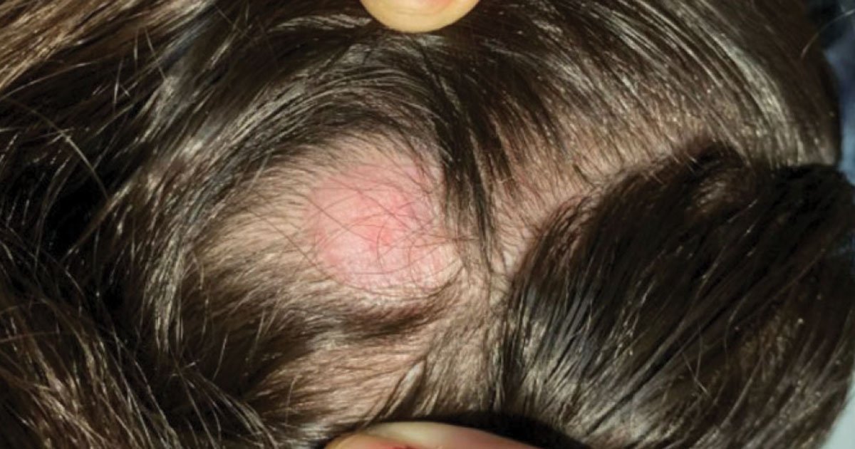
On examination, the affected person introduced to the emergency division with the next important indicators: temperature of 36.8°C, coronary heart price of 119 beats per minute, blood strain of 105/67 mm Hg, respiratory price of 27 breaths per minute and oxygen saturation of 100% on room air. She was awake, alert and oriented to particular person, place and time. She had no ache. A quick bodily examination revealed a patch of alopecia on her occipital scalp (Determine 1).
Preliminary ocular examination revealed close to imaginative and prescient of 20/40 in the correct eye with no enchancment on pinhole and lightweight notion within the left eye with no enchancment on pinhole. IOP was 15 mm Hg in the correct eye and 9 mm Hg within the left eye. The suitable pupil was spherical and briskly reactive with no afferent pupillary defect, whereas the left pupil was spherical and briskly reactive with a 2+ afferent pupillary defect. Extraocular actions have been full in each eyes. Within the left eye, confrontation visible fields have been absent in all 4 quadrants together with lack of shade imaginative and prescient. Slit lamp biomicroscopy examination was most notable for cryptless irides in each eyes (Determine 2) with Shafer’s signal within the left eye.

Dilated fundus examination revealed the next notable findings (Determine 3). In the correct eye, the vitreous media was clear. The optic nerve had hint pallor with sharp margins and a cup-to-disc ratio of 0.2. The macula had a round, well-delineated space of hypopigmentation with staphylomatous look that seemingly corresponded to central macular atrophy with a dimension of roughly 1.5 disc diameters (Determine 3a). The blood vessels have been regular caliber, and the restricted peripheral view revealed a power retinal detachment with in depth retinal pigment epithelium degeneration according to a myopic fundus. Conversely, within the left eye, the vitreous confirmed pigment cells and floaters. The optic nerve had hint pallor with sharp margins. The macula was indifferent from the retina and had a big atrophic macular gap. Vessels have been skinny with no apparent bleeding. Periphery revealed a complete macula-off retinal detachment with fastened folds (Determine 3b). There have been no different breaks on scleral depressed fundus examination.

Preliminary workup
Arriving at an correct analysis may be aided with the usage of numerous imaging modalities. To additional consider this pediatric bilateral retinal detachment (power in the correct eye and acute within the left eye) from the dilated fundus examination, a previous OCT of the macula was reviewed. OCT macula of the correct eye confirmed important macular atrophy with absent foveal pits and subfoveal scarring; equally, OCT macula of the left eye confirmed lack of foveal contour and macular atrophy (Determine 4). Fundus pictures of each eyes was additionally captured on the time of the preliminary encounter and highlighted the aforementioned findings from the dilated examination (Determine 3).

What’s your analysis?
See reply beneath.
Bilateral retinal detachment
With regard to the affected person’s chief grievance of acute imaginative and prescient loss within the left eye with bilateral retinal detachment discovered on ocular examination, it is very important maintain a broad differential in these circumstances.


The differential for a pediatric bilateral retinal detachment begins with consideration of the most typical causes of retinal detachment, together with trauma, myopia, prior ocular surgical procedure and historical past of retinopathy of prematurity. The affected person didn’t have a historical past of trauma, prior ocular surgical procedure or ROP. She was, nevertheless, a pathologic myope at –16 D in each eyes. It’s also essential to think about an infectious trigger on the differential. As an illustration, toxoplasmosis is the most typical reason behind posterior uveitis in youngsters, which leads to 6% of pediatric retinal detachments annually. Nonetheless, this affected person had regular vitals and labs with no febrile or infectious indicators on bodily or ocular examination. Subsequent, oncologic tumors and inflammatory causes have been thought of as a result of retinoblastomas and choroidal hemangiomas are attainable causes of retinal detachments in youngsters. Nonetheless, dilated fundus examination and OCT for this affected person didn’t reveal indicators of mass lesions or irritation comparable to uveitis, vitritis or vasculitis. The final however most essential class thought of was congenital vitreoretinopathies. This can be a group of genetic issues that manifests with congenital abnormalities of the vitreous and retina and predisposes pediatric sufferers to rhegmatogenous retinal detachments. Examples of congenital vitreoretinopathies embody Stickler syndrome, Wagner syndrome, snowflake vitreoretinal degeneration and Knobloch syndrome. Given this affected person’s historical past of younger age, household and private histories of pathologic myopia, cryptless irides on anterior section examination, macular atrophy and bilateral retinal detachment, congenital vitreoretinopathies have been highest on the differential.
Additional workup
A genetics seek the advice of was ordered to guage for a suspected congenital vitreoretinopathy. The genetics group accomplished a complete analysis of the affected person’s private, delivery and developmental historical past. Pertinent positives included a historical past of sort 1 diabetes, onset of lowering imaginative and prescient bilaterally on the age of two years and a patch of alopecia on her occipital scalp. She had no developmental or studying disabilities. A radical bodily examination confirmed no dysmorphic facial options, hypermobility, joint issues, rashes or birthmarks. Her echocardiogram was inside regular limits with no septal defects. A renal and bladder ultrasound was regular, excluding hydronephrosis. Neurosurgery was consulted to rule out occipital cranium defects, and endocrinology was consulted to additional optimize her diabetes. Provided that this affected person had bilateral retinal detachments within the setting of early onset myopia, craniofacial findings (occipital alopecia), anterior section abnormalities (cryptless irides) and systemic findings (sort 1 diabetes), the genetic and ophthalmology groups have been suspicious of a connective tissue dysfunction.
The differential was additional narrowed to connective tissue issues that lead to congenital vitreoretinopathies: Marfan syndrome, Ehlers-Danlos syndrome, Stickler syndrome and Knobloch syndrome. A genetic panel check was despatched by way of a saliva pattern, which revealed a defect within the COL18A1 gene.
Analysis
The affected person’s analysis was autosomal recessive Knobloch syndrome with a optimistic COL18A1 defect.
Administration
The remedy for this affected person included retinal surgical procedure for rhegmatogenous retinal detachment restore bilaterally and shut monitoring with pediatric ophthalmology. She underwent a pars plana vitrectomy, membrane peel, endolaser at 360°, placement of a 41 scleral buckle band tied with a 70 sleeve, and infusion of 5,000 centistoke silicone oil within the left eye with an skilled retinal surgeon. Postoperatively, the affected person recovered appropriately with no issues. Visible acuity within the left eye improved from gentle notion preoperatively to counting fingers at 4 ft on the postoperative 1-week follow-up and to twenty/200 on the 6-month follow-up. For the power retinal detachment found in the correct eye, the affected person underwent a pars plana vitrectomy, endolaser at 360°, cryotherapy, and placement of a 41 scleral buckle band tied with a 70 sleeve. Visible acuity in the correct eye improved from 20/400 preoperatively to twenty/150 on the 6-month follow-up.
Dialogue
That is an attention-grabbing pediatric case of Knobloch syndrome, which is a situation related to imaginative and prescient loss secondary to retinal detachments, occipital cranium defects and typically genitourinary abnormalities. Defects within the COL18A1 gene result in dysfunction of sort 18 collagen, which is crucial for the integrity of epithelial basement membranes in connective tissues all through the physique. Kids with Knobloch usually have bodily manifestations and extreme imaginative and prescient decline earlier than the age of 10 years.
Knobloch pathogenesis begins with a defect within the COL18A1 gene that produces faulty sort 18 collagen, leading to weak basement membranes. Irregular basement membranes disrupt the structural integrity of connective tissues, notably within the retina, cornea, mind and kidneys. Disrupted integrity of the basement membrane results in dysfunctional tissues that can’t keep intact and powerful sufficient to hold out their respective capabilities. For instance, the weakened epithelial basement membrane in retinal tissue results in macular hypoplasia, atrophy and finally a macular gap, together with the affected person on this case. Within the mind, this situation could current as occipital cranium defects, hydrocephalus and cerebral atrophy. Within the anterior eye section, sufferers could develop corneal dystrophies, and within the kidneys, it will possibly result in hydronephrosis. These bodily findings finally trigger lifelong debilitating medical signs comparable to imaginative and prescient loss, systemic endocrine and renal situations, and infrequently cognitive delays.
Sufferers with Knobloch syndrome have excessive ranges of vitreoretinal degeneration and near-universal occurrences of bilateral retinal detachments. It’s thought that disruption in vitreous composition and lack of structural integrity in epithelial basement membranes from dysfunctional sort 18 collagen is a key contributor to extreme visible loss in these sufferers. Vitreous is without doubt one of the many connective tissues within the physique. It’s recognized that sort 2 collagen is widespread in cartilage and vitreous. Nonetheless, analysis exhibits that sort 18 collagen can also be essential for anchoring vitreous collagen fibrils to the interior limiting membrane. Due to this fact, the vitreoretinopathy seen in Knobloch is characterised as early vitreous liquefaction, veils of vitreous collagen condensation and perivascular lattice within the peripheral retina. These adjustments improve the chance for a retinal detachment.
A typical triad seen in Knobloch syndrome with childish onset is vitreoretinal degeneration with retinal detachment, excessive myopia and occipital findings (starting from alopecia to encephalocele). If a affected person presents with this triad of signs, there ought to be a excessive diploma of suspicion for a genetic dysfunction and therefore a low threshold for genetic testing. Nonetheless, not all sufferers with Knobloch will current with the basic triad. Knobloch syndrome may also produce a various number of ocular signs together with band-shaped keratopathy, persistent fetal vasculature, cryptless irides, macular atrophy, atrophic macular holes, pale optic discs and absent foveal pits. Systemic manifestations could embody congenital hydronephrosis, hypermobile joints, midline occipital bone defects or alopecia on the posterior scalp, cognitive decline, seizures and even ventricular dilation.
Visible prognosis for sufferers with Knobloch syndrome is poor and requires lifelong shut ophthalmologic monitoring. Extreme, progressive, irreversible imaginative and prescient decline or loss is inevitable. Virtually all sufferers can have a bilateral retinal detachment earlier than the age of 10 years. Nonetheless, most sufferers might be able to reside common lives with low imaginative and prescient aids and strict precautionary efforts. Shut neurosurgery and nephrology follow-up may additionally be required within the setting of systemic abnormalities.
Remedy for these sufferers contains surgical and supportive interventions. Sufferers require low imaginative and prescient companies, up to date glasses and strict avoidance of contact sports activities. Genetic counseling and parental schooling are additionally essential so that folks are conscious of genetic dangers of passing the situation to future offspring and are finest geared up to help their little one at school and at house. Sufferers ought to bear a vitrectomy with scleral buckle and silicone oil tamponade if a retinal detachment happens. If a affected person is identified with Knobloch syndrome earlier than a retinal detachment, then prophylactic surgical procedure to keep away from retinal detachment formation is really useful.
Retinal detachments within the pediatric inhabitants account for 3.2% to six.6% of all retinal detachments. A noteworthy discovering in sufferers with Knobloch is that they generally have macular hole-related retinal detachments. A current research by Alzaben and colleagues investigated this distinctive type of retinal detachments in sufferers with Knobloch syndrome. This single-center retrospective research included 50 Knobloch sufferers from 1983 to 2023, and the researchers studied the speed, traits and outcomes of rhegmatogenous retinal detachments in these sufferers. They discovered that 74% of sufferers who have been clinically identified with Knobloch primarily based on bodily examination have been discovered to have a confirmatory gene defect. This signifies the position of the medical examination and historical past taking for early analysis and intervention in these sufferers, particularly if genetic testing is delayed or denied. Forty-eight eyes (48%) developed a retinal detachment with a imply age of 6.5 years, and of these with retinal detachments, 33% concerned a macular gap. By way of surgical remedy, the very best single-surgery success price was a mix vitrectomy with adjunct scleral buckle and silicone oil tamponade.
Scientific course continued
Two years after her bilateral vitrectomy, the affected person returned to the retina clinic with worsening imaginative and prescient within the left eye. She was discovered to have a dense cataract seemingly resulting from her earlier vitrectomy. The affected person underwent cataract surgical procedure within the left eye with symptom decision. Six months later, she returned to the retina clinic with recurrence of visible decline within the left eye. A dense posterior capsular opacification was noticed within the left eye, requiring in-clinic YAG laser capsulotomy. The affected person’s visible acuity efficiently returned to her postoperative baseline of 20/200 within the left eye. She continues to be adopted intently by pediatric and retina specialists.
- References:
- Alzaben KA, et al. Ophthalmol Retina. 2024;doi:10.1016/j.oret.2024.03.020.
- Islam Y, et al. https://www.aao.org/schooling/disease-review/pediatric-retinal-detachments. Revealed Sept. 28, 2021. Accessed Could 13, 2025.
- Kim LA, et al. Knobloch syndrome. https://eyewiki.org/Knobloch_Syndrome. Revealed Oct. 8, 2024. Accessed Could 13, 2025.
- Le Goff MM, et al. Eye (Lond). 2008;doi:10.1038/eye.2008.21.
- O’Neill MJF. Knobloch syndrome 1; KNO1. https://www.omim.org/clinicalSynopsis/267750. Up to date June 4, 2020. Accessed Could 15, 2025.
- Sebag J. Vitreous pathobiology and anomalous posterior vitreous detachment. https://entokey.com/vitreous-pathobiology-and-anomalous-posterior-vitreous-detachment/#R62-V3-39. Revealed July 11, 2016. Accessed Could 13, 2025.
- Sevcik Ok, et al. Stickler syndrome. https://EyeRounds.org/circumstances/320-stickler-syndrome.htm. Revealed Jan. 20, 2022. Accessed Could 13, 2025.
- Thau A, et al. Ophthalmic Surg Lasers Imaging Retina. 2019;doi:10.3928/23258160-20190806-13.
- Tozer KR, et al. Vitreous and developmental vitreoretinopathies. https://www.vmrinstitute.com/wp-content/uploads/2013/10/Hartnett-Ch-3.pdf. Revealed June 6, 2013. Accessed Could 13, 2025.
- Vasilyeva T, et al. Genes (Basel). 2024;doi:10.3390/genes15101295.
- For extra data:
- Edited by William W. Binotti, MD, and Julia Ernst, MD, PhD, of New England Eye Middle, Tufts College Faculty of Medication. They are often reached at william.binotti@tuftsmedicine.org and julia.ernst@tuftsmedicine.org.