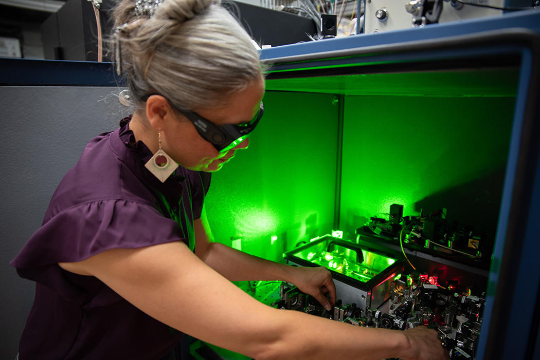
imaged using the platform described in the new paper in Nature Communications. Credit: Lingyan Shi lab, UC San Diego")
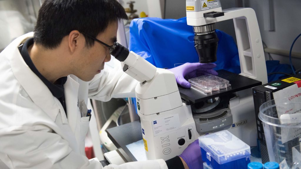
Scientists from the College of California San Diego and Washington College Faculty of Medication in St. Louis have developed a strong new technique to look inside kidney tissues—without having to stain or injury the tissue samples. The brand new method, referred to as label-free multimodal optical biopsy, captures detailed, 3D photos of kidney samples and divulges early indicators of illness usually missed by conventional strategies.
Revealed in Nature Communications, the research introduces a brand new imaging system that makes use of lasers to detect essential adjustments within the kidney’s construction and chemistry. This contains figuring out extra fats buildup, protein alterations, and tissue scarring—key warning indicators of diabetic kidney illness, one of many main causes of kidney failure worldwide.
“Consider it as a high-tech digicam that may ‘see’ inside tissue with out disrupting cells or requiring stains,” stated Lingyan Shi Ph.D., co-corresponding writer and professor within the Shu Chien-Gene Lay Division of Bioengineering at UC San Diego. “It provides us a extra full image of what is taking place contained in the kidney—and does it in 3D.”
Sanjay Jain MD Ph.D., co-corresponding writer and professor of medication, pathology and pediatrics at Washington College Faculty of Medication in St. Louis added, “This expertise has monumental potential to enhance how we diagnose and monitor kidney illness, particularly in its early phases when intervention may be handiest. It gives us clues to what changes or diversifications are taking place within the cells in diabetes and associated metabolic problems within the kidney.”
Anthony (Tony) A. Fung Ph.D. is the research’s first writer and a former bioengineering doctoral pupil and postdoctoral researcher in Shi’s bioengineering lab on the UC San Diego Jacobs Faculty of Engineering and at present a postdoctoral researcher at Yale College. Fung led the event and integration of the imaging platform and information evaluation pipeline used on this research whereas at UC San Diego.
In contrast to conventional biopsy strategies, which require a number of serial sections with chemical stains, co-registration post-processing, and dangers bodily slicing defects, this label-free strategy preserves the pattern whereas delivering wealthy, informative photos that mirror underlying biology. When tissue availability is sparse, sustaining the pliability for downstream processing is a key benefit of non-destructive label-free imaging.
To additional improve its impression, the workforce integrated synthetic intelligence to mechanically detect indicators of illness inside the imaging information. This functionality may sooner or later assist clinicians diagnose kidney issues extra rapidly and precisely, and personalize therapy plans for every affected person.
The researchers hope this expertise is not going to solely enhance early detection, monitoring, and therapy of kidney illness but additionally discover functions in learning different illnesses the place metabolism and tissue construction are essential.
Extra data:
Anthony A. Fung et al, Label-free multimodal optical biopsy reveals biomolecular and morphological options of diabetic kidney tissue in 2D and 3D, Nature Communications (2025). DOI: 10.1038/s41467-025-59163-w
Quotation:
A clearer take a look at diabetic kidney illness by way of new optical imaging expertise (2025, Could 19)
retrieved 19 Could 2025
from https://medicalxpress.com/information/2025-05-clearer-diabetic-kidney-disease-optical.html
This doc is topic to copyright. Other than any honest dealing for the aim of personal research or analysis, no
half could also be reproduced with out the written permission. The content material is supplied for data functions solely.